Articles
- Page Path
- HOME > J Educ Eval Health Prof > Volume 13; 2016 > Article
-
Research article
The student experience of applied equivalence-based instruction for neuroanatomy teaching -
W. James Greville1*
 , Simon Dymond2,3
 , Philip M. Newton4
-
DOI: https://doi.org/10.3352/jeehp.2016.13.32
Published online: September 13, 2016
1Department of Psychology, Aberystwyth University, Aberystwyth, United Kingdom
2Department of Psychology, Swansea University, Swansea, United Kingdom
3Department of Psychology, Reykjavík University, Reykjavík, Iceland
4Research in Health Professions Education, Swansea University Medical School, Swansea, United Kingdom
- *Corresponding email: wjg3@aber.ac.uk
Copyright © 2016, Korea Health Personnel Licensing Examination Institute
This is an open-access article distributed under the terms of the Creative Commons Attribution License, which permits unrestricted use, distribution, and reproduction in any medium, provided the original work is properly cited.
Abstract
-
Purpose
- Esoteric jargon and technical language are potential barriers to the teaching of science and medicine. Effective teaching strategies which address these barriers are desirable. Here, we created and evaluated the effectiveness of stand-alone ‘equivalence-based instruction’ (EBI) learning resources wherein the teaching of a small number of direct relationships between stimuli (e.g., anatomical regions, their function, and pathology) results in the learning of higher numbers of untaught relationships.
-
Methods
- We used a pre and post test design to assess students’ learning of the relations. Resources were evaluated by students for perceived usefulness and confidence in the topic. Three versions of the resources were designed, to explore learning parameters such as the number of stimulus classes and the number of relationships within these classes.
-
Results
- We show that use of EBI resulted in demonstrable learning of material that had not been directly taught. The resources were well received by students, even when the quantity of material to be learned was high. There was a strong desire for more EBI-based teaching. The findings are discussed in the context of an ongoing debate surrounding ‘rote’ vs. ‘deep’ learning, and the need to balance this debate with considerations of cognitive load and esoteric jargon routinely encountered during the study of medicine.
-
Conclusion
- These standalone EBI resources were an effective, efficient and well-received method for teaching neuroanatomy to medical students. The approach may be of benefit to other subjects with abundant technical jargon, science and other areas of medicine.
- Consider some basic neuroanatomical terms: ‘substantia nigra,’ ‘hippocampus,’ ‘pons,’ and ‘cerebellum,’ These structures all perform essential functions; medical students should be able to identify them, describe what they do and the consequences of damage to them. However, these names are derived from Latin or Greek words which are, at best, only distantly related to the medical relevance of these brain regions or to everyday language. The abundance of esoteric jargon has long been suggested as a reason for the persistence of rote learning strategies in medical education [1].
- Jargon is a source of cognitive load [2]. The cognitive load theory of education is based upon research showing that human working memory capacity is extremely limited and easily overloaded, but that the processing of new information through working memory is essential for that information to be integrated into existing knowledge (i.e., learned) [3]. Thus there is a need to limit extraneous sources of cognitive load and focus on relevant information only [4,5]. Unfortunately the esoteric language of medicine can quickly cause working memory to become overloaded.
- Equivalence-based instruction (EBI) is an instructional method based upon the theory of ‘stimulus equivalence’ [6]. A defining feature of EBI is that the subject learns more than that which is directly taught, and thus the efficiency of instruction is maximized. For example, if a student is taught a relationship between stimulus, ‘A’ (e.g., the words ‘substantia nigra’) and stimulus ‘B’ (e.g., a picture of the substantia nigra), and then taught a relationship between ‘A’ and a different stimulus ‘C’ (e.g., ‘the brain region lost in Parkinson’s disease’), then they will learn, without being directly taught, a relationship between ‘B’ and ‘C’; when shown pictures of unlabelled brain regions and asked to identify which of them is lost in Parkinson’s disease, they will correctly select the picture of the ‘substantia nigra.’ This outcome is called ‘stimulus equivalence’ [7]. The addition of a single additional taught relationship (e.g., between ‘A’ and stimulus ‘D’ ‘produces dopamine’) causes an exponential increase in the number of relationships learned without direct teaching (i.e., B=D, C=D). These interrelated stimuli (A to D) are then recognized as equivalent.
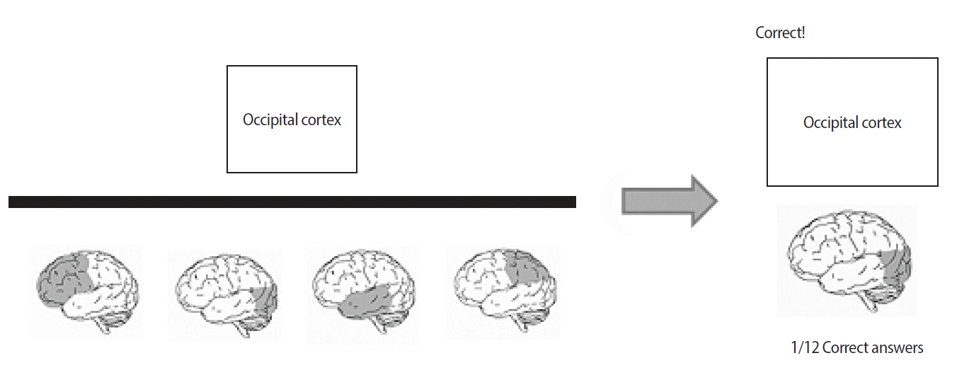
- These relationships are quickly learned using a ‘match-to-sample’ (MTS) paradigm [6] consisting of learning events in which a participant is presented with an initial stimulus, known as ‘the sample.’ A number of comparison stimuli are then presented, from which the participant must the select the one which best ‘matches the sample’ (e.g., as shown in Fig. 1, the words ‘occipital cortex’ could be the sample, with pictures of the different cortical regions as comparison stimuli). Feedback may be given following each selection, to facilitate learning.
- A handful of studies have experimentally demonstrated that EBI is effective as a strategy in ‘higher education.’ Fienup et al. [8] used EBI in an experimental laboratory setting to teach some basic neuroanatomy to small (4 student) groups of volunteer psychology students, while Pytte and Fienup [9] showed EBI was effective within a large-group instructor-led lecture-based setting.
- Here, we evaluated the student experience of applied EBI using standalone resources streamlined to optimize the learning experience, and determine whether increasing the size and complexity of resources affected this experience. We focused on neuroanatomy in order to build on studies from Fienup et al. [8]. Also, it has been proposed that medical students/trainees will experience ‘neurophobia’: a lack of confidence and understanding in neuroanatomy/neuroscience, caused, in part, by the complexity of these topics and the (perceived) quality/quantity of the teaching received [10,11]. Learning resources aimed at improving confidence and understanding of neuroanatomy are therefore desirable.
Introduction
- Three resources were designed, in a consecutive manner. Relations are shown in Table 1 and Table 2. Participants were medical students at Swansea University, from both years 1 and 2. A power analysis using G*Power (GPower Software Inc., Kiel, Germany) [12] recommended a sample size of 45 (one-tailed with power 0.95, effect size 0.5 and α 0.05); our sample sizes were 43, 24, and 39 in resources 1, 2, and 3, respectively. Students completed the resource either in timetabled 25 minutes sessions or in their own time. Resources were advertised by email and in teaching sessions. No incentives were given for completing the resources, besides the learning experience itself. Resources were created using E-Prime ver. 1.3 from Psychology Software Tools Inc. (Pittsburgh, PA, USA) and were available on Windows 7 PCs with 19-inch widescreen LCD displays. Images used in Table 1 were adapted from an image in the public domain. Images were used in Table 2 under the terms of creative commons license CC-BY-SA-2.1-JP issued by the creator: BodyParts3D, © The Database Center for Life Science. Items included in the resource were selected following consultation and pilot testing with medical students interested in the subject.
- Research design and resource structure
- We utilized a quasi-experimental pretest-postest design. Resources comprised three main phases; (1) pre-test of untaught relations and self-reported confidence in the subject, (2) learning of taught relations, and (3) post-test of (same) untaught relations and self-reported confidence in the subject. Each consisted of a series of learning events in MTS format. An example is shown in Fig. 1. To orient participants to the layout, detailed on-screen instructions were given followed by a simple MTS example: the word ‘blue’ was shown as the sample above four squares; blue, red, green, and yellow; participants had to select the blue square.
- Pre-learning test of untaught relations
- This tested participants baseline level of knowledge of the ‘equivalence’ relationships that would not be directly taught in the learning phase (e.g., B=C, B=D, and C=D). This is a subset of the total untaught relations, ignoring symmetrical variants (C=B, D=B, and D=C), and symmetrical variants of taught relations (B=A, C=A, and D=A). As our focus was on the student experience we felt it unnecessary to provide an exhaustive, repetitive test of all relations. For advanced learners, competence on a subset of equivalence relations implies competence on their symmetrical variants [8].
- Participants were first asked “How would you rate your current level of confidence with regard to your knowledge of neuroanatomy? 1=weak, 3=average, 5=strong” on a 5-point Likert scale. They then began the pre-test. Each untaught equivalence relation was presented once only, in random order, with no feedback and a brief (1 second) blanking of the screen between. Upon completion, participants were told their score. Participants scoring 80%+ were advised that their knowledge of neuroanatomy appeared to already be quite high and thus they may not benefit from the resource, but that they were free to continue should they wish. All participants elected to proceed.
- Learning of taught relations
- This phase comprised repeated MTS learning blocks of all those relations with stimulus ‘A’ as the root (A=B, A=C, A= D, etc.), for each brain region in the resource. Each block contained all the different combinations of category and stimulus class being taught (A1B1, A2B2, A3B3, A4B4, A1C1, etc.) once only, in random order, before beginning a new block. Feedback was provided; if the incorrect stimulus was selected, the screen cleared and the word ‘incorrect’ was displayed in red for 3 seconds. If the choice was correct, the word ‘correct!’ was displayed in blue, together with the sample stimulus and the target stimulus, again for 3 seconds (as shown in Fig. 1). After each question participants were told how many correct answers they had achieved thus far. Blocks repeated until participants attained a required criterion of consecutive correct responses (see below).
- Post-learning test of untaught relations
- This was a repeat of the pre-test and confidence rating. Students were also then asked further feedback questions; firstly, “how useful did you find this learning resource?” on a five-point Likert scale with ‘not at all useful’ and ‘extremely useful’ attached as descriptive labels to points 1 and 5, respectively; secondly, “would you like more resources structured in this way?” with yes or no as response options. Further feedback comments were collected on paper or via email.
- Specific details of individual resources
- Resource 1 piloted EBI-based neuroanatomy teaching using four of five categories (A-D) shown in Table 1, thus there were 12 taught relations (learning phase) (A=B, A=C, and A= D×4 cortical regions) and 12 untaught relations in pre/post-test (B=C, B=D, and C=D×4 regions). Criterion for completing the learning phase was all 12 taught relations answered correctly.
- Resource 2 (Table 1) included 16 (4 relations×4 regions) taught relations per block. Criterion for completing the learning phase was 16 consecutive correct answers. There were 24 untaught relations in the pre/post test phases (B=C, B=D, B=E, C=D, C=E, and D=E relations×4 regions).
- Resource 3 covered three relations for eight different neuroanatomical structures, giving 24 taught relations (Table 2). Criterion for completing the learning phase was 22/24 correct within a given block with 24 untaught relations in the test phases (B=C, B=D, and C=D relations, 8 structures).
- Statistical analyses
- Wilcoxon matched-pairs signed rank tests were used to compare mean scores across participants on the pre-test and post-test, both for the percentage of correct answers on the untaught relations test, and confidence ratings. One-sample Wilcoxon signed rank tests were used to compare overall usefulness ratings against the ‘indifference’ midpoint of 3.
- Ethical approval
- It was obtained, prior to data collection, from the joint ethics committee of the Colleges of Medicine and Health and Human Sciences at Swansea University, United Kingdom.
Methods
- Twelve taught relations
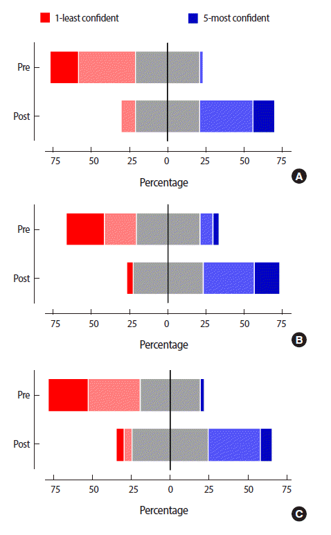
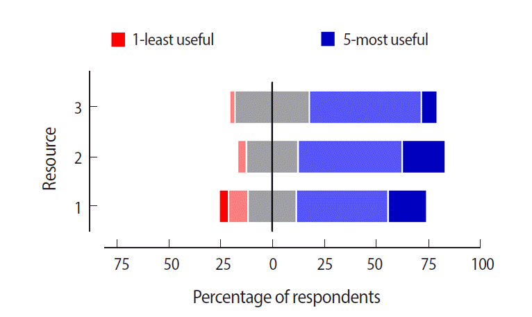
- Results are summarized in Table 3. Mean score on post-test (94%) was significantly higher than on pre-test (75%), W=-727, P<0.0001. 25/43 participants (57%) scored 100% on post-test, indicating a ceiling effect. There was also a significant improvement in participants confidence, W=-682, P< 0.0001 (Fig. 2). Students rated the resource as useful (Fig. 3), Z=3.282, P<0.005. 88% (38/43) indicated they would like additional resources in this format.
- Qualitative student feedback was positive (Table 4), with a desire for more of these resources being a recurring theme. Given that dissatisfied service users are more likely to provide negative feedback than satisfied users are to provide positive feedback, this was particularly encouraging. Improvements were also suggested. The small number of negative comments highlighted the repetitive feature of the EBI approach.
- Sixteen taught relations
- Results are shown in Table 3. Scores on the pretest of untaught relations were lower compared to resource 1, presumably due to the additional untaught relations. Despite the sample size being underpowered for this resource, a significant learning effect was again observed; mean score of 91% on post-test being significantly higher than pre-test (46%), W=-274, P<0.0001. 16/24 participants (66.7%) scored 100% in the post-test, again suggesting a ceiling effect. Confidence again improved (Fig. 2), W=-132, P<0.0016, and students found the resource useful, Z=3.586, P<0.001 (Fig. 3). 92% (22/24) of students indicated that they would like to see more resources in this format.
- Twenty-four taught relations
- Results are summarized in Table 3. Scores in the post-test (93%) were again significantly higher than on the pre-test (64%), W=-780, P<0.0001. 8/39 (21%) showed maximum scores of 100%. Confidence in knowledge of the topic was again improved, W=-553, P<0 .0001 (Fig. 2) and usefulness rating was again significantly higher than the indifference midpoint of 3, Z=4.435, P<0.001.
- This resource included an additional evaluation question to probe the student experience of using EBI versus other methods. Participants were asked “please take a moment to think about the amount of time you have spent in completing the resource, what you have learned, and if this has improved your confidence in the subject of neuroanatomy. Now think about other ways you might have spent this time to study the same topic, such as reading a textbook. Overall, how well-spent do you feel your time has been in completing the resource compared to other instructional methods?” This question was presented immediately after the ‘usefulness’ question. Responses were on a 5-point Likert scale, where 1 was designated as ‘not as useful as learning from a textbook,’ 3 was designated as ‘just as useful as…’ and 5 was ‘more useful than….’ 76% of students favored the resource over a textbook (42.1% selecting ‘4’ on the Likert scale and 34.2% selecting ‘5’). Overall rating was significantly higher than the midpoint of the scale (‘3,’ ‘just as useful’), Z=4.518, P<0.001.
- Comparison of the student experience across all three resources
- Resources 1-3 contained increasing levels of complexity, yet completion of all resources was associated with statistically significant increases in confidence (Fig. 3) with no changes in ratings of ‘usefulness’; direct comparison of the rated ‘usefulness’ of each resource using a Kruskal-Wallis test revealed no significant difference, H=1.208, P=0.55.
Results
- In the three studies presented here, medical students using standalone EBI-based resources to learn neuroanatomy reported a highly positive learning experience, and a significant increase in their confidence even when faced with learning a considerable volume of learning. Increasing the numbers of relations did not diminish the positive learning experience.
- Improved confidence and engagement are perhaps the most salient of our findings, relating directly to the proposed causes of ‘neurophobia.’ A student having a positive learning experience, paired with a demonstrable outcome of having learned the name, location and basic function of major neuroanatomical regions in approximately 25 minutes, may be less daunted by more complex neuroanatomy. Structuring learning resources in this way also signposts students to the fundamental scaffolding of the neuroanatomy under study, something which might not be so obvious from an alternate mode of study (e.g., reading a textbook). Indeed, 76% of our participants rated even the most complex learning resource as more useful than a textbook when considering time invested.
- Our findings support the claim of Pytte and Fienup [9] that EBI may be effectively implemented within a natural teaching environment at a university. Pytte and Fienup [9]’s classroom-based study appears to require a substantial investment of time, effort and planning to teach some very basic neuroanatomy. Our simpler and more cost-effective approach may best utilize the benefits of EBI through self-contained standalone online resources.
- Despite these positive findings, EBI is not appropriate for all contexts in medical education; healthcare professionals clearly rely on much more than simple structure-function-pathology relationships. A small number of our participants expressed the opinion that the resources represent rote learning, possibly due to their repetitive nature. It is a widely held view that rote learning is an undesirable educational strategy [13]. A persistence of rote learning in medical education has been thought to arise, at least in part, through the existence of esoteric jargon and factual overload [1]. Rote learning is not universally regarded as negative; from the perspective of cognitive load theory it has been suggested that rote learning may facilitate the committing of core knowledge and skills to ‘automaticity,’ thereby freeing up space in the limited working memory capacity for deeper levels of thinking [14]. The approach demonstrated here, while possibly repetitive or even rote, ensures that esoteric jargon is placed directly into the relevant structure-function relationship using an extremely efficient and active form of learning, rather than traditional rote-learning or mnemonics.
- There are a number of limitations to the interpretation of these data, most of which result from our taking the EBI paradigm out of the laboratory and into real classrooms. For example, students were not required to complete the resource, or to allow us to collect their responses, all of which were anonymous. Thus the positive student experience may be inflated by self-selection, compounded by the fact that our sample sizes were quite low. Further research would be helpful to determine whether the positive findings generated in this context generalize to other concepts and disciplines. It would also be useful to determine whether the information learned during EBI can be retained for a longer period of time, or to directly test how it compares when directly tested against other, more traditional teaching methods for neuroanatomy.
Discussion
-
Conflict of interest
No potential conflict of interest relevant to this article was reported.
-
Funding
This work was supported by a grant from the Swansea Academy of Learning and Teaching to Philip M. Newton and Simon Dymond.
Article information
Supplementary materials


The eight stimulus classes pertained to various neuroanatomical structures, while the categories described either structure name, its anatomical location on a diagram, a particular function of the structure, and a result of pathological damage to the structure. In each case the image shown is one slice of a total twelve frames, the composite of which created a rotating animated GIF.
| Resource |
Relations |
Untaught relation test (%) |
||||
|---|---|---|---|---|---|---|
| Taught | Untaught | No. | Pre | Post | More? | |
| 1 | 12 | 12 | 43 | 75.3 | 94.2 | 88 |
| 2 | 16 | 24 | 24 | 46.0 | 91.3 | 92 |
| 3 | 24 | 24 | 39 | 64.1 | 93.1 | 97 |
- 1. Pandey P, Zimitat C. Medical students’ learning of anatomy: memorisation, understanding and visualisation. Med Educ 2007;41:7-14. http://dx.doi.org/10.1111/j.1365-2929.2006.02643.x ArticlePubMed
- 2. Sweller J. Element interactivity and intrinsic, extraneous, and germane cognitive load. Educ Psychol Rev 2010;22:123-138. http://dx.doi.org/10.1007/s10648-010-9128-5 Article
- 3. Dehn MJ. Working memory and academic learning: assessment and intervention. Hoboken (NJ): John Wiley & Sons; 2011.
- 4. De Jong T. Cognitive load theory, educational research, and instructional design: some food for thought. Instr Sci 2010;38:105-134. http://dx.doi.org/10.1007/s11251-009-9110-0 Article
- 5. Mayer RE. Applying the science of learning to medical education. Med Educ 2010;44:543-549. http://dx.doi.org/10.1111/j.1365-2923.2010.03624.x ArticlePubMed
- 6. Sidman M. Symmetry and equivalence relations in behavior. Cogn Stud 2008;15:322-332. http://dx.doi.org/10.11225/jcss.15.322
- 7. Dymond S, Roche B. Advances in relational frame theory: research and application. Oakland (CA): New Harbinger; 2013.
- 8. Fienup DM, Covey DP, Critchfield TS. Teaching brain-behavior relations economically with stimulus equivalence technology. J Appl Behav Anal 2010;43:19-33. http://dx.doi.org/10.1901/jaba.2010.43-19 ArticlePubMedPMC
- 9. Pytte CL, Fienup DM. Using equivalence-based instruction to increase efficiency in teaching neuroanatomy. J Undergrad Neurosci Educ 2012;10:A125-131. PubMedPMC
- 10. Zinchuk AV, Flanagan EP, Tubridy NJ, Miller WA, McCullough LD. Attitudes of US medical trainees towards neurology education: “Neurophobia”: a global issue. BMC Med Educ 2010;10:49. http://dx.doi.org/10.1186/1472-6920-10-49 ArticlePubMedPMCPDF
- 11. Flanagan E, Walsh C, Tubridy N. ‘Neurophobia’: attitudes of medical students and doctors in Ireland to neurological teaching. Eur J Neurol 2007;14:1109-1112. http://dx.doi.org/10.1111/j.1468-1331.2007.01911.x ArticlePubMed
- 12. Faul F, Erdfelder E, Lang AG, Buchner A. G*Power 3: a flexible statistical power analysis program for the social, behavioral, and biomedical sciences. Behav Res Methods 2007;39:175-191. http://dx.doi.org/10.3758/BF03193146 ArticlePubMed
- 13. Mayer RE. Applying the science of learning. Boston (MA): Pearson/Allyn & Bacon; 2011.
- 14. Hattie J, Yates G. Visible learning and the science of how we learn Abingdon: Routledge; 2014.
References
Figure & Data
References
Citations

- Twelve tips for teaching neuroanatomy, from the medical students’ perspective
Sanskrithi Sravanam, Chloë Jacklin, Eoghan McNelis, Kwan Wai Fung, Lucy Xu
Medical Teacher.2023; 45(5): 466. CrossRef - Comparing two equivalence‐based instruction protocols and self‐study for teaching logical fallacies to college students
Emily E. Gallant, Kenneth F. Reeve, Sharon A. Reeve, Jason C. Vladescu, April N. Kisamore
Behavioral Interventions.2021; 36(2): 434. CrossRef - Neuroanatomy, the Achille’s Heel of Medical Students. A Systematic Analysis of Educational Strategies for the Teaching of Neuroanatomy
Maria Alessandra Sotgiu, Vittorio Mazzarello, Pasquale Bandiera, Roberto Madeddu, Andrea Montella, Bernard Moxham
Anatomical Sciences Education.2020; 13(1): 107. CrossRef - Developing and Implementing Emergent Responding Training Systems With Available and Low-Cost Computer-Based Learning Tools: Some Best Practices and a Tutorial
Bryan J. Blair, Lesley A. Shawler
Behavior Analysis in Practice.2020; 13(2): 509. CrossRef - Sidman Goes to College: A Meta-Analysis of Equivalence-Based Instruction in Higher Education
Julia Brodsky, Daniel M. Fienup
Perspectives on Behavior Science.2018; 41(1): 95. CrossRef - Tools and resources for neuroanatomy education: a systematic review
M. Arantes, J. Arantes, M. A. Ferreira
BMC Medical Education.2018;[Epub] CrossRef - Corrigendum: Misplacement of images in a table including the structure of the cerebral cortex
Journal of Educational Evaluation for Health Professions.2018; 15: 12. CrossRef

 KHPLEI


 PubReader
 ePub Link
 Cite
Cite


